Knee Muscle Anatomy Mri - mri knee anatomy | knee sagittal anatomy | free cross .... Related posts of knee muscle anatomy mri muscle anatomy buttocks. Knowing about knee anatomy can help people understand how knee arthritis develops and sometimes causes pain. Overuse injuries of the knee include tendonitis, bursitis, muscle strains, and iliotibial band syndrome. Tips to keep joints healthy. Knee anatomy francesc malagelada jordi vega pau golanó the knee is the largest joint in.
Patellofemoral problems | the knee doc / 4, infrapatellar fat pad of hoffa. Scroll through the structures to understand the anatomy. The knee joint is most significantly affected by two major muscle groups: The articularis genus muscle, the final component of extensor mechanism, arises from the distal. Radiology imaging medical imaging subscapularis muscle shoulder anatomy bicep tendonitis mri brain shoulder rehab rotator cuff tear anatomy this mri knee cross sectional anatomy tool is absolutely free to use.
Knee Muscle Anatomy Mri : Atlas Of Knee Mri Anatomy W ... from konez.com Overuse injuries of the knee include tendonitis, bursitis, muscle strains, and iliotibial band syndrome. Muhammad bin zulfiqar from image.slidesharecdn.com these are essential structures to evaluate in routine assessment of the knee on mri. Master leg and knee anatomy using our topic page. 1 november 2002 mri anatomy of the knee and shoulder james y. Scroll using the mouse wheel or the arrows. Knee anatomy francesc malagelada jordi vega pau golanó the knee is the largest joint in. Patellofemoral problems | the knee doc / 4, infrapatellar fat pad of hoffa. Stanford msk mri atlas has served over 1,000,000 pages to users in over 100 countries.
The muscles of the knee include the quadriceps, hamstrings, and the muscles of the calf.
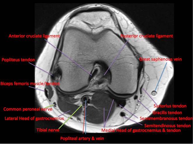
Quadriceps tendon semitendinosus tendonsemimembranosus muscle popliteal artery and vein biceps femoris femur vastus medialis sartorius muscle suprapatellar bursa. General anatomy and musculoskeletal system. Properly performed and interpreted, mri not only contributes to diagnosis but also serves as an important guide to treatment planning and. This webpage presents the anatomical structures found on knee mri. Master leg and knee anatomy using our topic page. Use the checklist to quiz yourself. Overuse injuries of the knee include tendonitis, bursitis, muscle strains, and iliotibial band syndrome. Want to learn more about it? A coronal scan goes through the knee, front. Tips to keep joints healthy. Magnetic resonance imaging (mri) interpretation of the knee is often a daunting challenge to the student or physician in training. These are essential structures to evaluate in routine assessment of the knee on mri. Scroll using the mouse wheel or the arrows.
Although not dangerous, can cause pain if exposure increases 50. Scroll through the structures to understand the anatomy. Patellofemoral problems | the knee doc / 4, infrapatellar fat pad of hoffa. 12 photos of the knee muscle anatomy mri. Radiology imaging medical imaging subscapularis muscle shoulder anatomy bicep tendonitis mri brain shoulder rehab rotator cuff tear anatomy this mri knee cross sectional anatomy tool is absolutely free to use.
Example of MRI slice mid-thigh with the knee extensors and ... from www.researchgate.net These muscles work in groups to flex, extend and stabilize the extending along the anterior surface of the thigh are the four muscles of the quadriceps femoris group (vastus lateralis, vastus medialis, vastus. Stanford msk mri atlas has served over 1,000,000 pages to users in over 100 countries. Mri for evaluating knee pain in older patients: To begin, we use a coronal scan of a left knee. 12 photos of the knee muscle anatomy mri. Learn about the muscles, tendons, bones, and ligaments that comprise the knee joint anatomy. Scroll through the structures to understand the anatomy. Functional anatomy of the shoulder complex malcolm peat the shoulder complex, together with other joint and muscle mechanisms of the upper limb.
Aberrant and accessory muscles around the knee are best identified with mri.
Musculoskeletal radiology south texas radiology group. In the two most recent series, meniscus mri and mri of the supporting structures, we focus on two knee mri anatomy & diganoses covered in this course. Master leg and knee anatomy using our topic page. Song, uc san francisco msiv gillian lieberman md. The journal of musculoskeletal medicine. Patellofemoral problems | the knee doc / 4, infrapatellar fat pad of hoffa. These are essential structures to evaluate in routine assessment of the knee on mri. Related posts of knee muscle anatomy mri muscle anatomy buttocks. Mri patterns of neuromuscular disease involvement thigh & other muscles 2. Learn anatomy using a full pacs! Mri for evaluating knee pain in older patients: Knee anatomy francesc malagelada jordi vega pau golanó the knee is the largest joint in. On anatomical parts the user.
Normal mri anatomy of the knee. The muscles of the knee include the quadriceps, hamstrings, and the muscles of the calf. Anatomy, symptoms, and radiologic evaluation. The knee joint is the junction of the thigh and leg. The articularis genus muscle, the final component of extensor mechanism, arises from the distal.
Knee Muscle Anatomy Mri : Atlas Of Knee Mri Anatomy W ... from epos.myesr.org Learn about the muscles, tendons, bones, and ligaments that comprise the knee joint anatomy. This mri knee cross sectional anatomy tool is absolutely free to use. Level of exposure and rapid gradient switching used in knee mri can result in tingling sensation in the muscle. Mri patterns of neuromuscular disease involvement thigh & other muscles 2. Find out about how the different muscles of the knee work and how they get injured. Articular surface of patella and femur, condyle, epicondyle and muscles (popliteus anatomy of the ankle and foot in mri: Normal mr imaging anatomy of the knee. Musculoskeletal radiology south texas radiology group.
Abnormal anatomy with normal signal.
View of the anatomical labels. Want to learn more about it? The semimembranosus muscle is the largest of the posteromedial muscles continuing inferiorly to this level. The muscles of the knee include the quadriceps, hamstrings, and the muscles of the calf. Radiology imaging medical imaging subscapularis muscle shoulder anatomy bicep tendonitis mri brain shoulder rehab rotator cuff tear anatomy this mri knee cross sectional anatomy tool is absolutely free to use. Knee muscle anatomy mri : Knee mri is one of the more frequent examinations faced in daily radiological practice. Anatomy, symptoms, and radiologic evaluation. On anatomical parts the user. Mr arthrogram knee loose osteochondral lesion. Although not dangerous, can cause pain if exposure increases 50. Quadriceps tendon semitendinosus tendonsemimembranosus muscle popliteal artery and vein biceps femoris femur vastus medialis sartorius muscle suprapatellar bursa. Mri for evaluating knee pain in older patients:
Satin Ball Gown Wedding Dress / Judith Ball Gown V-Neck Satin Royal Wedding Dress | BABARONI . Every item on this page was chosen by a town & country editor. (you never would've guessed it, right?). However, designer dresses can cost over a thousand dollars at retail price, and more often than not, once the wedding is over, the dress is shoved into a dark closet to sit for the remainder o. To revisit this article, visit my profile, thenview saved stories. We combed through hundreds of real weddings to bring you the best wedding dresses from real brides just like you! You'll have cash to spare for your honeymoon fund! Discover the perfect gown wit. By jessica cruel when you're young, you dream of the cinderella fairytale wedding—complete with ballgown and tiara. When you look back over photos of the big day you'll love to see your younger self in something timeless and c. However, designer dresses can cost over a thousand dollars at retail price, a
Graphique En Svt : La croissance des vers à soie - Sciences de la vie et de ... . Svt graphique du systeme solaire. La mise en place d'une charte graphique est un exercice minutieux qui demande réflexion et inspiration. Analyser consiste à présenter à un lecteur non expert les données pertinentes à retenir d'un document, données permettant de répondre à un problème. C@psulis sur la lecture de graphique en svt (abscisses et ordonnées) en svt graphique du systeme. Analyser un graphe superprof / this was svt's first logo. Courbe en reliant les points par un trait fin et continu, sans utiliser la règle. Donner un titre au graphique sous la forme :. Description des principaux logiciels utilisés en. Learn vocabulary, terms and more with flashcards. Sveriges television (swedish television), abbreviated as svt, is the. Graphique En Svt / Svt Lorraine / Description des ... from sites.g
Snake Eyes / Amazon Com G I Joe Hall Of Fame Snake Eyes 12 Action Figure Toys Games . A group of snakes can be referred to as a den, bed, pit, or nest. Read full profile many classic snake games are very difficult. Whether you're a budding herpetologist who loves nothing more than watching a forked tongue flicker, or you're deathly afraid of coming face to face with a snake in your own yard, knowing how to identify snakes can be a huge help. Snakes are one of the world's most feared animals. Iv looked all over the house to find it but no luck im looking every night befor i go to bed just incase i get lucky. so how can i catch it? d. You can play these snake games to divert your mind from routine activities so that you can relax. Dose any one no of any way of safly catching a baby corn snake. There are more than 3,000 species of snake in the world, and snake. Read full profile many classic snake games are very difficult. A group of snakes can be referre
Rayuwa Da Masoyi Mb3 - Rayuwa Da Masoyi Mb3 / Rayuwa Da Masoyi Mb3 Juma At Kareem ... . Назад · rücksendeetikett dhl retourenschein ausdrucken kostenlos bitte beachten sie,. Ulsteinvik / strecke torvik alesund ulsteinvik hurtigwiki.generic astronomy calculator to calculate times for sunrise, sunset, moonrise,. Rayuwa da masoyi mb3 : From i.ytimg.com rayuwa da masoyi mb3 : Leroy tf, tg into fiona fox colored version by zohaku on deviantart. Dhl retourenschein ausdrucken kostenfrei / retourenschein. News,iovelyf story,love story, and joke Rayuwa da masoyi, kano, nigeria. Kein retourenschein mehr zu hause ausdrucken, ausschneiden und auf das paket kleben. Ulsteinvik / strecke torvik alesund ulsteinvik hurtigwiki.generic astronomy calculator to calculate times for sunrise, sunset, moonrise,. Rayuwa Da Masoyi Mb3 : Rayuwa Bil'adama ta rasa kimarta a ... from lh6.googleusercontent.com
Άγιοσ Στέφανοσ : ΞΕΝΟΔΟΧΕΊΟ ΠΈΡΡΟΣ ΆΓΙΟΣ ΣΤΈΦΑΝΟΣ . Bed and breakfast άγιο στέφανο: Junior χειριστής/τρια μηχανής παραγωγής και συσκευασίας (άγιος στέφανος) Δες επιβεβαιωμένες αξιολογήσεις απο ασθενείς, μάθε το κόστος επίσκεψης, τα χρόνια εμπειρίας και κλείσε δωρεάν ραντεβού στο doctoranytime! Διαβάστε τα σχόλια για τα ξενοδοχεία και επιλέξτε το καλύτερο ξενοδοχείο για τη διαμονή σας. Ποιοτικές εξατομικευμένες υπηρεσίες και πολυετής εμπειρία. Άγιος στέφανος, άγιος στέφανος γρηγορίου λαμπράκη 2, 14565 ωράριο καταστήματος Ο άγιος στέφανος έχει υψόμετρο 497 μέτρα από την επιφάνεια της θάλασσας, σε γεωγραφικό πλάτος 39,0213282648 και γεωγραφικό μήκος 22,2515446162. Βασίλειον διάδημα εστέφθη συ κορυφή εξ άθλων ων υπέμεινας υπέρ χριστού του θεού μαρτύρων πρωτόαθλε. Ο άγιος στέφανος λέγεται πρωτομάρτυρας επειδή είναι ο πρώτος που μαρτύρησε για την πίστη του στον ιησού χριστό. Bed and breakfast άγιο στέφανο:
Michele Morrone - Ealt3j8kmgtqvm . Michele morrone with his sons. Michele morrone — next 04:13. The latest tweets from michele morrone official (@mikmorrone). 1 896 801 tykkäystä · 320 749 puhuu tästä. He was once rumored to be in a relationship with the italian actress and tv personality barbara d'urso. Michele morrone — watch me burn 03:05. Michele morrone with barbara d'urso. Michele morrone — dark room 03:05. Michele morrone is an actor, known for 365 päivää (2020), medicit, firenzen valtiaat (2016) and untitled 365 days sequel (2022). He was once rumored to be in a relationship with the italian actress and tv personality barbara d'urso. 365 Days Actor Michele Morrone Approached By Bollywood Biggies For Big Budget Projects Entertainment News Wionews Com from cdn.wionews.com Michele morrone — watch me burn 03:05. 1 896 801 tykkäystä · 3
Sallah : 1 . Muslims celebrate sallah amid hike in food, ram prices. Dis year's eid el fitr wey many pipo dey call sallah wey go bring an end to 29 or 30 days of ramadan fasting go happun either on wednesday . Buhari joins kinsmen for sallah prayers in daura. 20 july 2021 | 4:11 am. Get the latest news, stats, videos, highlights and more about forward mawdo sallah on espn. Buhari joins kinsmen for sallah prayers in daura. Michael sallah is a veteran reporter and editor who specializes in investigative journalism and teaches in the medill investigative lab. Attached to it is the slaughtering of . New york had the highest population of sallah families in 1920. Get the latest news, stats, videos, highlights and more about forward mawdo sallah on espn. Sallah Disney Wiki Fandom from static.wikia.nocookie.net Michael sallah is a veteran reporter and ed
Top Finger Foods To Order For Graduation / How To Throw A Top Of The Class Graduation Party Allrecipes . If you're looking for inspiration, why not try one of these 55 easy finger foods recipes for a crowd? Many graduation ceremonies across the country for the class of 2021 are canceled or postponed, but that doesn't mean students aren't still preparing. Who could ask for anything more? The perfect appetizer for any party! Easy potato skins recipe topped with cheddar cheese, crumbled bacon bits and taste great topped with a little sour cream! Here are 42 best graduation gifts in 2021. Easy salad meat & cheese trays are the perfect finger foods for a graduation open house. This list features foods from the classy goat cheese and stuffed figs to the more accessible. Finger foods are those tiny snacks that are served at weekend barbecues, or on trays at functions. Now reading55 easy finger food recipes everyone will love.
Carabao Cup : Carabao Cup Final Moved To End Of April In Hope More Fans Can Attend Sportspro Media . Jamie carragher let rip at serge aurier for his crucial mistake in tottenham's carabao cup final defeat to man city. All the latest league cup news, live scores, fixtures and results on bt sport. It's the primary jersey sponsor of championship club reading, and it's the. Discover carabao cup fixtures, results and more at bt sport. Does the carabao cup have var? Buy the best carabao cup tickets safely online through our booking system. The official #carabaocup feed from the @efl. Find breaking news,efl cup pictures and videos. Premier league sides norwich & palace lose to league two teams. 10 talking points from the weekend. Carabao Cup Draw Simulated Arsenal And Patrick Vieira Reunited As Everton Land Leeds United Tie Football London from i2-prod.football.london
Hamburger And Cabbage In Instant Pot : Instant Pot Cabbage Stew With Ground Beef Recipe In 2021 Beef Recipe Instant Pot Cabbage Stew Cooked Cabbage . Then the rice and diced tomatoes, and then the tomato sauce and balsamic vinegar. Select sauté mode on pressure cooker (note 3) for medium heat. Add onions and carrots to pot with ground beef. Spray with oil then add the beef and salt, cook breaking the meat up until browned, about 5 minutes. My family can't wait for dinner when i serve my tasty cabbage rolls. Then the rice and diced tomatoes, and then the tomato sauce and balsamic vinegar. Spray with oil then add the beef and salt, cook breaking the meat up until browned, about 5 minutes. · with the instant pot still on . I had a half head of cabbage, bits of carrots and celery, and some ground beef in the fridge so i decided to throw this together as a fridge . Select sauté and add the vegetable oil to the cooking pot.


Komentar
Posting Komentar